Acetabular Anteversion App
Acetabular version refers to the anteroposterior orientation of the acetabular opening relative to the true horizontal axis of the pelvis. The normal human acetabulum is anteverted in order to allow impingement free range of motion including flexion, adduction, and internal rotation. Abnormal acetabular version has been correlated with pathologic hip conditions including femoroacetabular impingement (FAI) and developmental hip dysplasia.The association between acetabular version and hip pain has been well established in recent years as a source of contributing to early hip osteoarthrosis and labral tears. Recognition and appropriate treatment of abnormal acetabular version is crucial to preventing irreversible damage to the hip


The normal human acetabulum is anteverted in order to allow impingement free range of motion including flexion, adduction, and internal rotation. Acetabular Abnormal acetabular version has been correlated with pathologic hip conditions including femoroacetabular impingement (FAI) and developmental hip dysplasia. Acetabular version refers to the anteroposterior orientation ofthe acetabular opening relative to the true horizontal axis ofthe pelvis.

The App is medical software aimed for orthopaedic surgeons, providing tools that allow doctors to: -Securely import medical images directly from the camera or stored photos. -By marking certain points in a simple standard AP pelvic radiograph, geometric parameters are being calculated. The App computes -The data are printed over to screen so each case can easily assessed -Save the planned images, for later review or consultation.The measured values are compared by normal reference databa

The App is medical software aimed for orthopaedic surgeons, providing tools that allow doctors to: -Securely import medical images directly from the camera or stored photos. -By marking certain points in a simple standard AP pelvic radiograph, geometric parameters are being calculated. The App computes -The data are printed over to screen so each case can easily assessed -Save the planned images, for later review or consultation.The measured values are compared by normal reference databa

The normal human acetabulum is anteverted in order to allow impingement free range of motion including flexion, adduction, and internal rotation. Acetabular Abnormal acetabular version has been correlated with pathologic hip conditions including femoroacetabular impingement (FAI) and developmental hip dysplasia. Acetabular version refers to the anteroposterior orientation ofthe acetabular opening relative to the true horizontal axis ofthe pelvis.
Acetabular version is conventionally evaluated on CT scans but excessive radiation doses associated with routine use of computed tomography (CT). An objective radiographic tool which provides measurements comparable in accuracy to CT measurements has been developed by Dr. Hefti (Nomogram).
Tedious and time-consuming calculation has to be done in simple X-rays in order to calculate the acetabular anteversion. The primary goal of this App is to help determine radiographic values of acetabular anteversion in a practice in a blink of an eye and avoiding CT scans.
The App is medical software aimed for orthopaedic surgeons, providing tools that allow doctors to:
-Securely import medical images directly from the camera or stored photos.
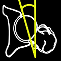
-By marking certain points in a simple standard AP pelvic radiograph, geometric parameters are being calculated. The App computes the acetabular anteversion based on a pelvic AP radiograph. The angle of anteversion is calculated through the formula according to Heftis nomogram. The acetabular orientation (anteversion/retroversion) is determine by the app by the measurement of the angles between the center of the femoral head and the anterior (φ) and posterior (φ’) acetabular rim.Once you choose correctly the anterior and posterior acetabular rims the app calculates the acetabular anteversion based on nomogram.
-The data are printed over to screen so each case can easily assessed
-Save the planned images, for later review or consultation.
All information received from the software output must be clinically reviewed regarding its plausibility before patient treatment! TheApp indicated for assisting healthcare professionals. Clinical judgment and experience are required to properly use the software.The software is not for primary image interpretation.
In a busy everyday practice, the examiner have to draw lines in X-rays or in clinical settings, this it is time consuming and cumbersome. Accessory instruments like protractors, hinged goniometers, well sharped pencils, calculators, rulers or even transparent papers must be available. The build in feature of the app, allows to calculate easily the version angle easily and help decide what could be considered normal or pathologic. This App is particular useful in clinical settings where you need a quick results without losing time. Please see tutorial videos at the developer’s web site www.orthopractis.com
Reference
Book. Pediatric Orthopaedics in Practice Friz Hefti ,Chapter 5 pelvis hips and thighs 5.3.2.1 Biomechanics of the hip- Springer.
Ηοw to measure with the App
The first thing is to load one image from your photo library or capture a photo from x-rays photos of a patient. By moving the transparent circular yellow template over the femoral head, trying to fit to a best-fit circle to the contour of the femoral head circumference. Once you have found the best fit by clicking the “point” button, the center of the femoral head is marked (C1). Next a dynamic circle appears with the C1 center point marked over the screen. The radius of the circle is changed dynamically and by moving the attached finger you try to find the best fit circle to the contour of the femoral head
Once you found the best fit you press the “point” button and the femoral head is marked with the circle with radius (R2). Next, you mark the intersection of the circle of femoral head with anterior acetabular rim and by clicking the button ‘point’ the point A3 appears. Similarly, you try to mark the intersection of the circle of femoral head with posterior acetabular rim and by pressing the button ‘point’ the A4 is printed. An orange vertical line is drawed from the femoral head center in order to secure the validation of measure by choosing A3, A4 in the correct equatorial side. According to Heftis nomogram the acetabular orientation (anteversion/retroversion) is determined with the app by measuring the angles φ and φ’ and a value is given.
In quick view, the reference points that you have to choose sequentially (manually) are shown below by the following order:
C1 → center of femoral head center
R2 → dynamic cycle radius of femoral head
A3 → intersection of radius of femoral head and anterior acetabular rim.
A4 → intersection of radius of femoral head and posterior acetabular rim.
Please see tutorial videos at the developer’s web site.
All information received from the software output must be clinically reviewed regarding its plausibility before patient treatment! TheApp indicated for assisting healthcare professionals. Clinical judgment and experience are required to properly use the software. The software is not for primary image interpretation.

